Core Lecture: Management of APL

Acute Promyelocytic Leukemia — diagnosis, risk stratification, and treatment

Ashwin Kishtagari, MBBS, MSCI

Division of Hematology/Oncology, Vanderbilt University Medical Center

2026-08-11

Outline

  1. Review of morphologic identification of APL
  2. Diagnosis and basic pathophysiology
  3. Risk stratification in APL
  4. Low-risk treatment — decoding the term “Lo-Coco”
  5. High-risk treatment
  6. Post-treatment monitoring and relapse treatment

The case

Case presentation

HPI

  • 31 y/o female with gum bleeding, night sweats, and 10 lb unintentional weight loss

Initial labs

  • WBC 6.4, Hgb 7.6, Plt 12
  • Differential shows 60% blasts
  • INR 1.6, PTT 45, Fibrinogen 106

Hypergranular promyelocytes on the peripheral smear

You page the path resident, who says it looks like APL. You order a STAT FISH AML panel for PML-RARA and start ATRA 22.5 mg/m² BID.

Epidemiology

  • 600–800 cases per year in the US
  • 5–10% of all acute myeloid leukemias
  • Median age at diagnosis 40 y (AML is 68 y)
  • Highly curable
  • Early death rate of 29% due to bleeding
  • Coagulopathy present in 80% of cases at presentation
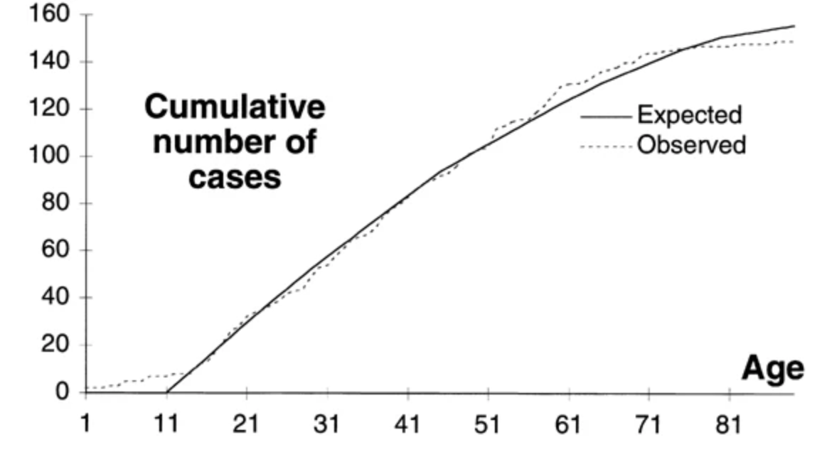
Cumulative number of APL cases by age

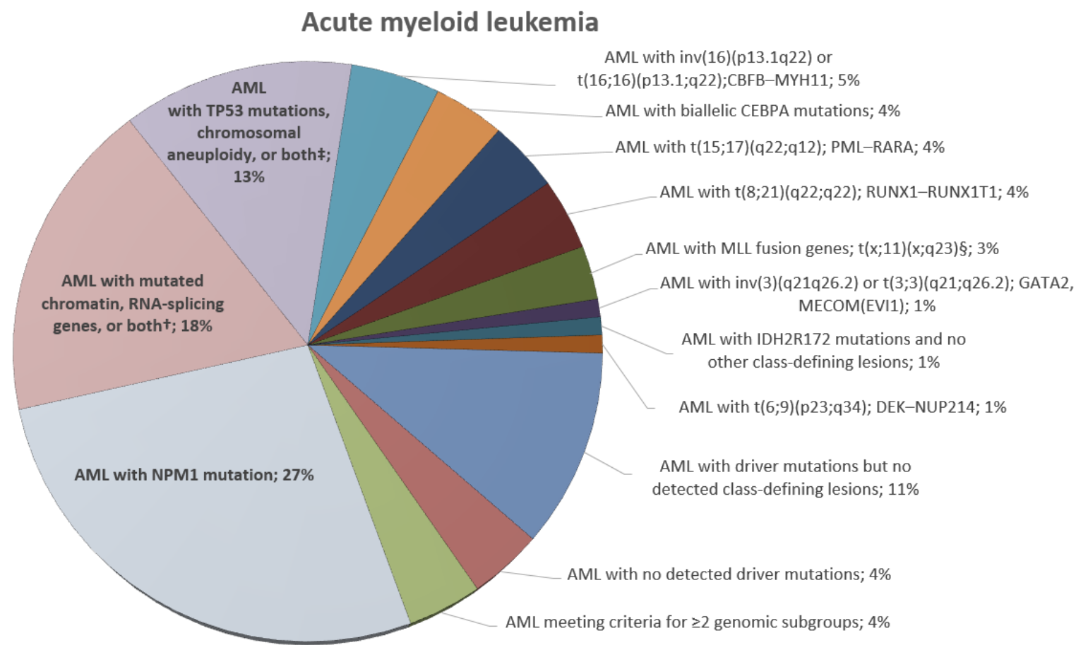
APL in the AML landscape

APL — t(15;17)/PML-RARA — is a small, genomically simple slice of the AML pie

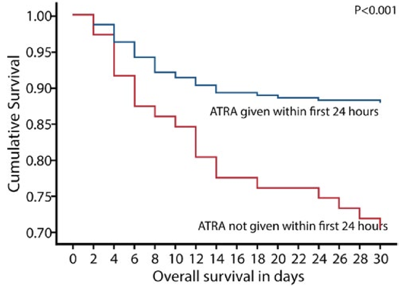
Early death is the enemy

Diagnosis and pathophysiology

APL diagnosis based on cytogenetics

  1. Suspect APL from the morphology and concomitant DIC
  2. Start ATRA 22.5 mg/m² BID on any suspicion — early death from coagulopathy is the threat
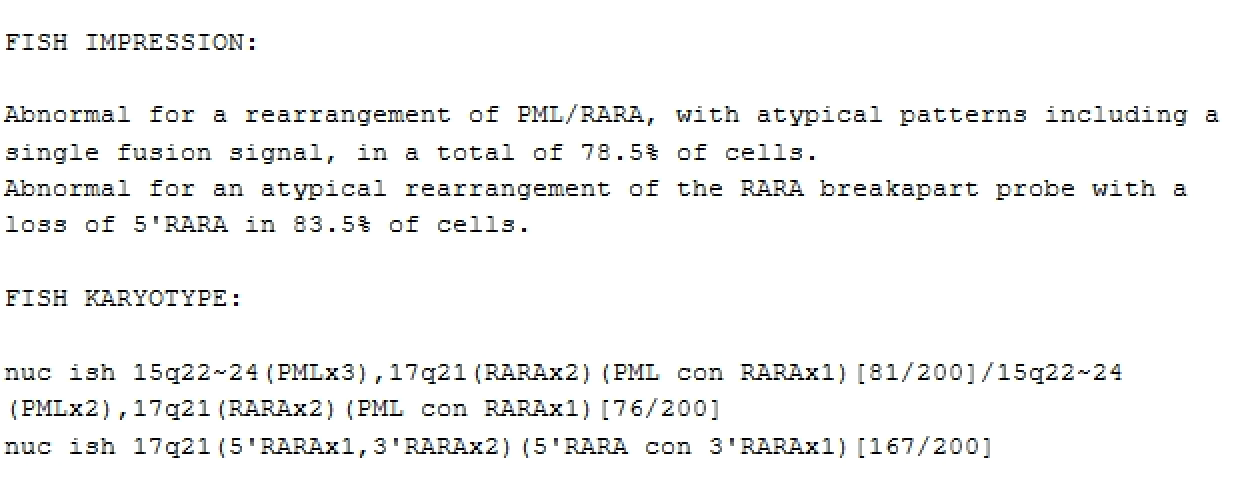
  3. Positive FISH or karyotype for the PML-RARA fusion gene → t(15;17), or
  4. Atypical translocations — but always involving 17
    • Chromosome 17 = RARA gene location
    • t(5;17), t(11;17), t(17;17)
  5. Much less genetic complexity than AML
    • RAS/FLT3 mutations occur but do not change risk stratification

APL morphology basics

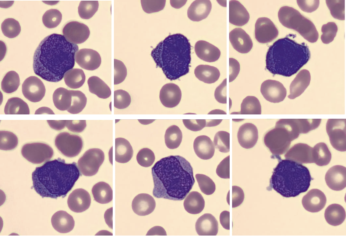
Hypergranular APL (60–70%)

  • Low WBC at presentation
  • Blasts can vary in size
  • Densely packed azurophilic granules and Auer rods
  • Lots of red-to-purple granules in the cytoplasm

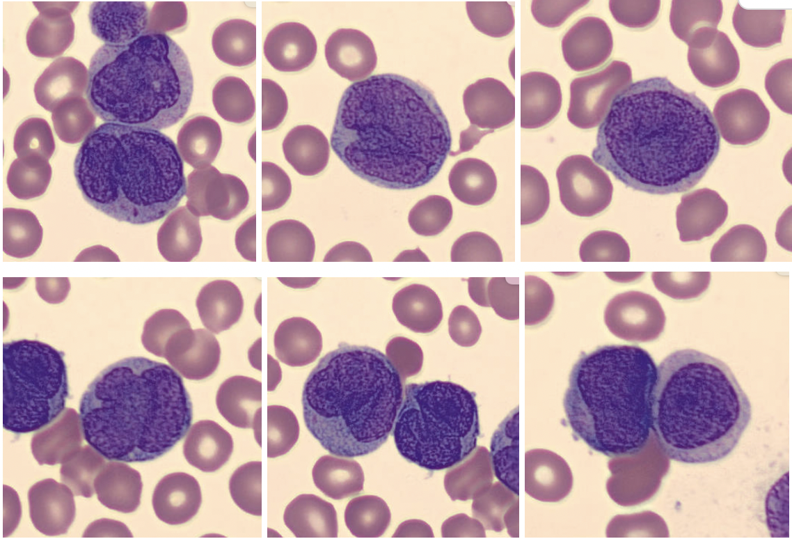
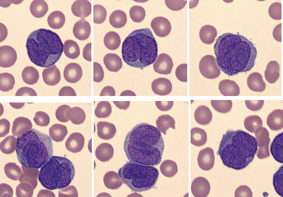
Hypogranular / microgranular variant

  • Leukocytosis
  • Associated with FLT3 mutations
  • Fewer, finer granules
  • Bilobed nuclei — “Venn diagram” or “dumbbell” shape

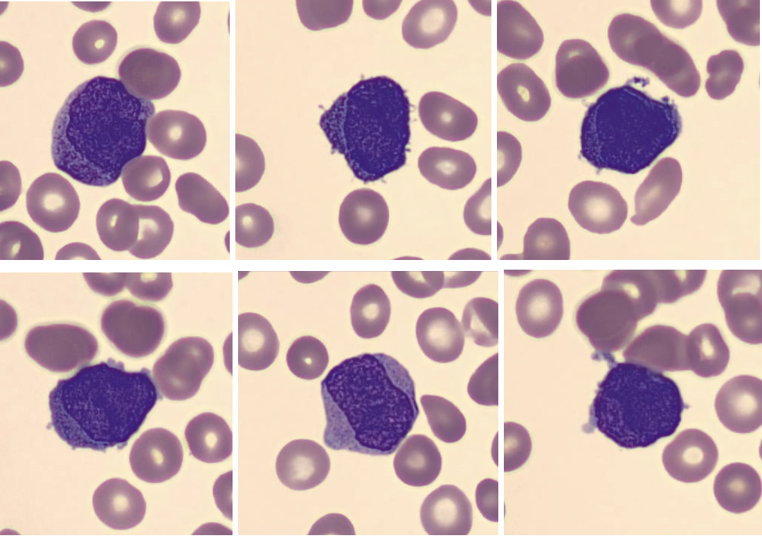
Hypergranular APL

Zoom in to see the purple granules filling the cytoplasm.

Hypogranular APL: bilobed nuclei
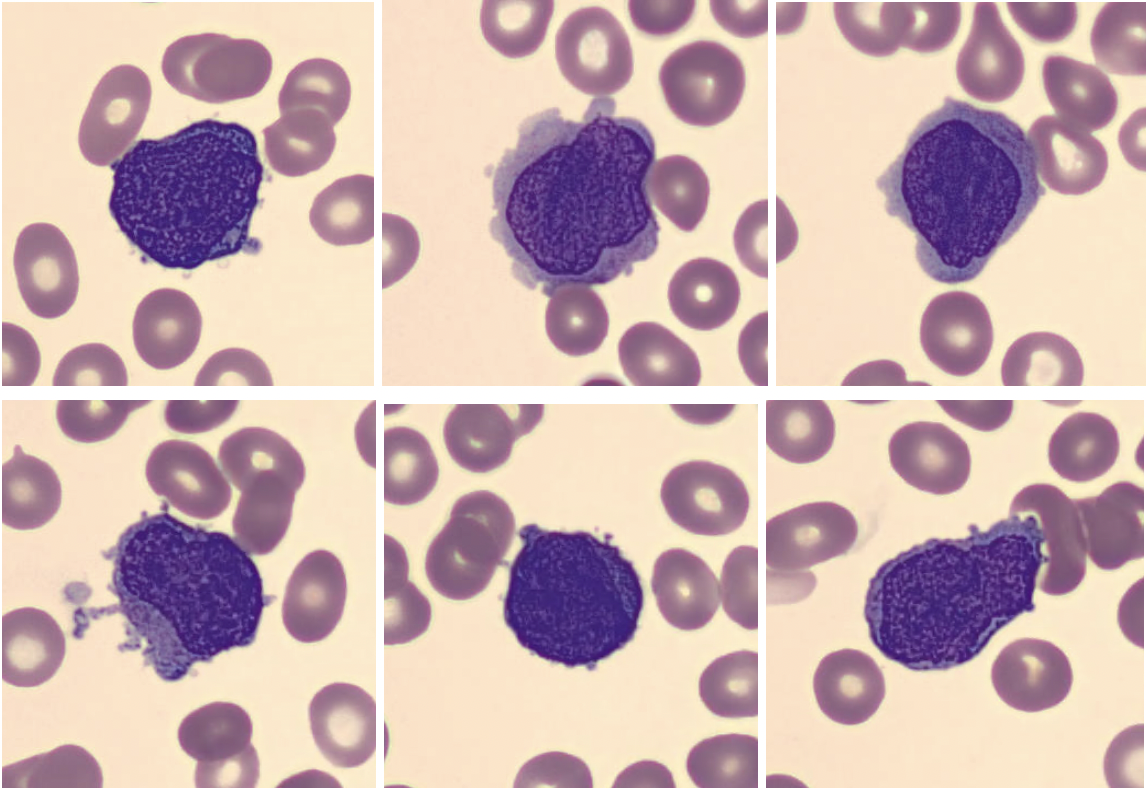
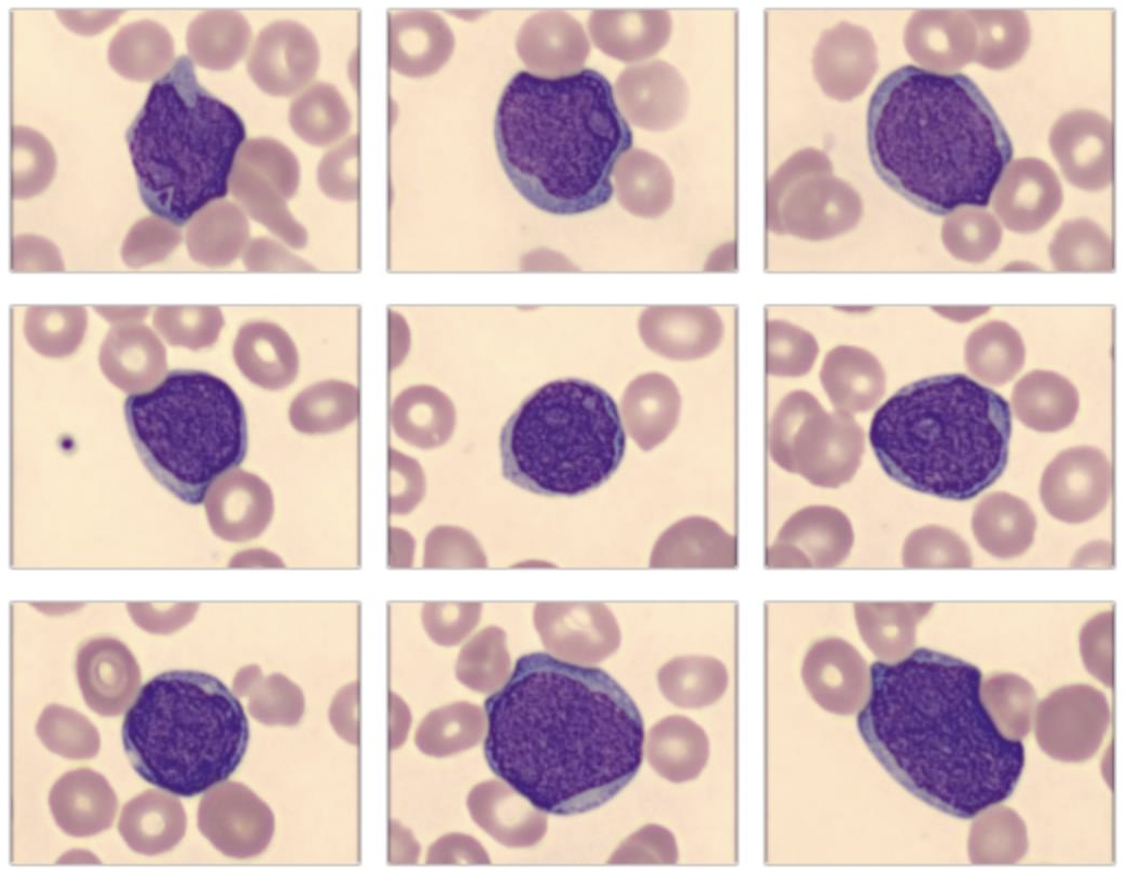
AML blast example — for contrast

  • High N:C ratio, much larger than background RBC
  • Clearly fewer granules, not as dark purple as hypergranular APL
  • No Venn diagram / bilobed nucleus

Boards flow cytometry for APL

Hypergranular

  • Increased side scatter → side scatter = granularity
  • CD33+, CD117+, MPO+
  • HLA-DR −, CD34 −

Hypogranular / microgranular variant

  • No increased side scatter
  • CD33+, CD34+
  • HLA-DR −

The constant across both: HLA-DR negative.

Our patient’s flow results

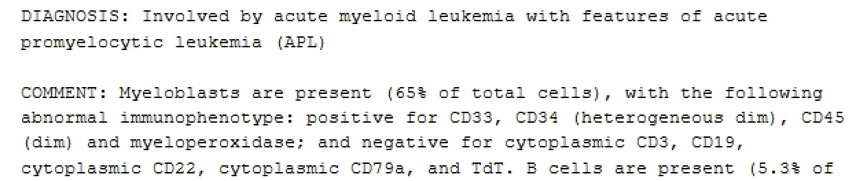
Not all patients follow the “typical pattern.” Here there is no comment on side scatter or HLA-DR — which is fine. Morphology makes you concerned, and PML-RARA makes the diagnosis.

Our patient’s FISH report… atypical?
What’s the deal with PML-RARA?

PML — chromosome 15

  • Promyelocytic leukemia gene; normally controls self-renewal
  • t(15;17) → self-renewal broken

RARA — chromosome 17

  • Retinoic acid receptor alpha; normally controls myeloid differentiation
  • Other partners possible in variant APL — t(x;17)

Cells can’t differentiate — stuck at the promyelocyte / blast stage.
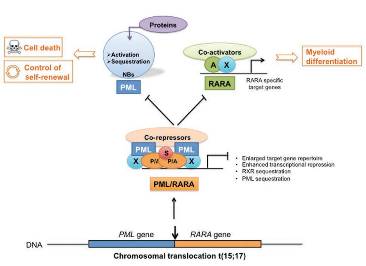
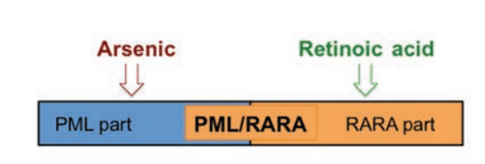
Why do we use ATRA and ATO?

ATRA = all-trans retinoic acid

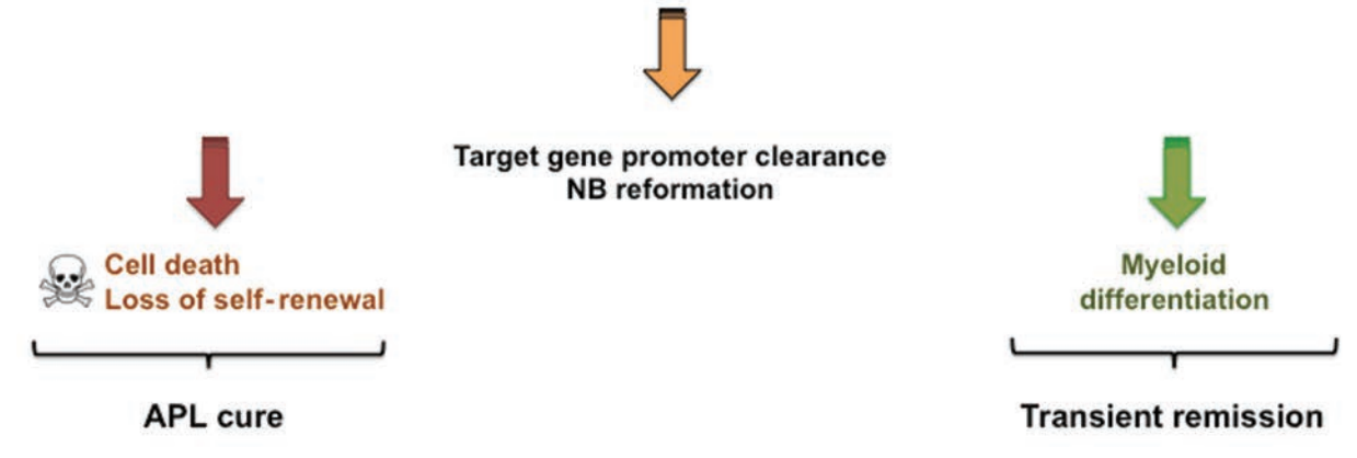
  • Prevents repressor activity of pathogenic RARA dimers → allows myeloid differentiation
  • WBC will rise as cells mature into neutrophils

ATO = arsenic trioxide

  • Leads to degradation of PML
  • Restores normal cell death and loss of self-renewal
Back to the case…

She had a bone marrow biopsy and was diagnosed with APL based on PML-RARA FISH and a karyotype with t(15;17).

Initial labs: WBC 6.4 · Hgb 7.6 · Plt 12 · 60% blasts · INR 1.6 · PTT 45 · Fibrinogen 106

Discussion

How often do you check DIC labs? Do you always use FFP for INR > 2?

  • Keep fibrinogen > ~150 → practically, 10 units of cryoprecipitate
  • Keep Plt > 50 → irradiated, to prevent transfusion-associated GVHD
  • Keep INR < 2 → can give vitamin K, but no evidence

Risk stratification

Case 1: APL, lower risk

Initial labs: WBC 6.4 · Hgb 7.6 · Plt 12 · 60% blasts · INR 1.6 · PTT 45 · Fibrinogen 106

Step 1 — Risk stratification: presenting WBC count

  • Low risk: WBC < 10
  • High risk: WBC > 10
    • Historically platelet count was used, but not currently

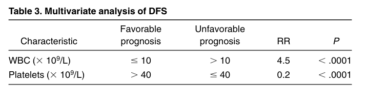
Where the Sanz score came from

  • Older study, pre-arsenic era — Plt <40 still incorporated
  • Patients got ATRA + anthracycline induction, then consolidation and maintenance
  • Primary endpoint: recurrence-free survival, not OS
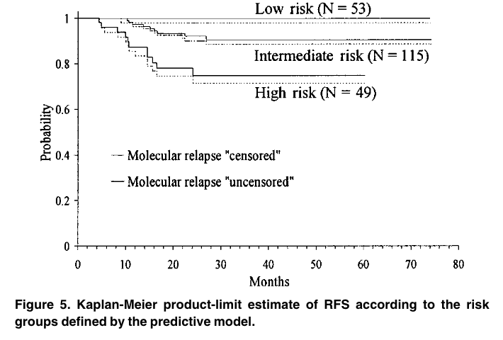
Sanz risk stratification for APL

  • Low risk: WBC <10, Plt >40
  • Intermediate risk: WBC <10, Plt ≤40
  • High risk: WBC >10

WBC >10 carries a far higher relative risk of relapse than Plt <40 — and the study predates arsenic. That is how the field ended up using WBC only for treatment decisions.

Low-risk treatment

Case 1: APL, low risk

Initial labs: WBC 6.4 · Hgb 7.6 · Plt 12 · 60% blasts · INR 1.6 · PTT 45 · Fibrinogen 106

Step 1 — Risk stratification

  • Low risk: presenting WBC < 10

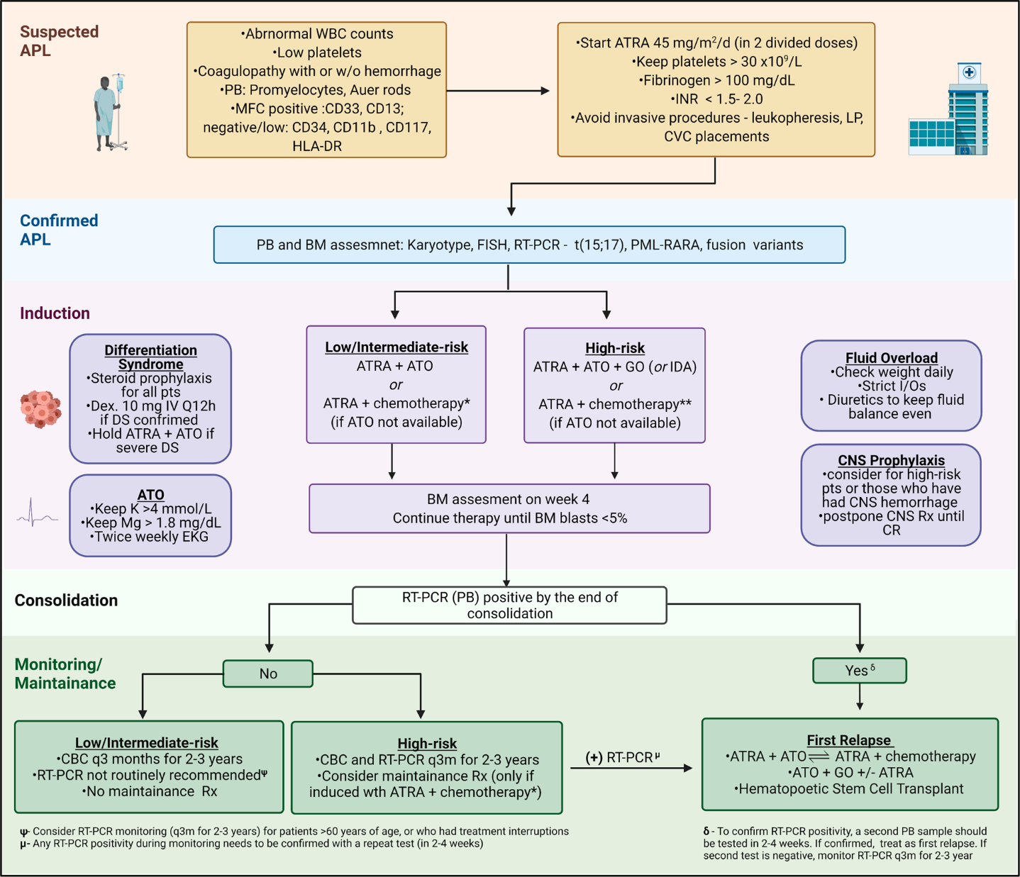
Step 2 — Initiate ATRA + ATO

  • ATRA 22.5 mg/m² PO BID → pediatric dosing differs
  • ATO 0.15 mg/kg IV daily over 2 hours

ATRA + ATO treatment monitoring

Step 2 — Initiate ATRA + ATO (low or intermediate risk)

  • ATRA 22.5 mg/m² PO BID · ATO 0.15 mg/kg IV daily over 2 hours

Step 3 — Treatment monitoring

  • LFTs at least 3× weekly
  • QTc 2× weekly — ATO causes QTc prolongation and arrhythmia; optimize K/Mg, avoid QTc-prolonging meds
  • Prednisone 0.5 mg/kg/day for differentiation syndrome prophylaxis — ATRA + ATO drives differentiation to mature cells, causing a SIRS picture

Discussion

Do you monitor LFTs more frequently up front? How do you handle a lengthening QTc during treatment?

Differentiation syndrome: fever + fluid

  • On-target adverse effect associated with differentiation therapy
  • Cytokine release and systemic inflammation that can result in shock, third spacing, pleural infiltrates, respiratory failure, and even death!
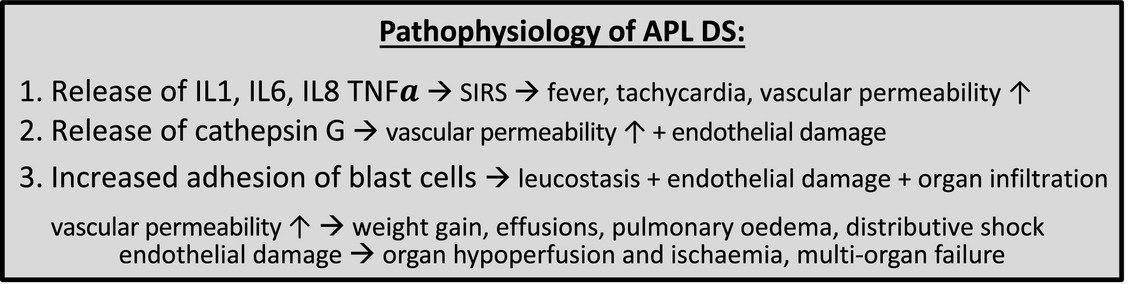
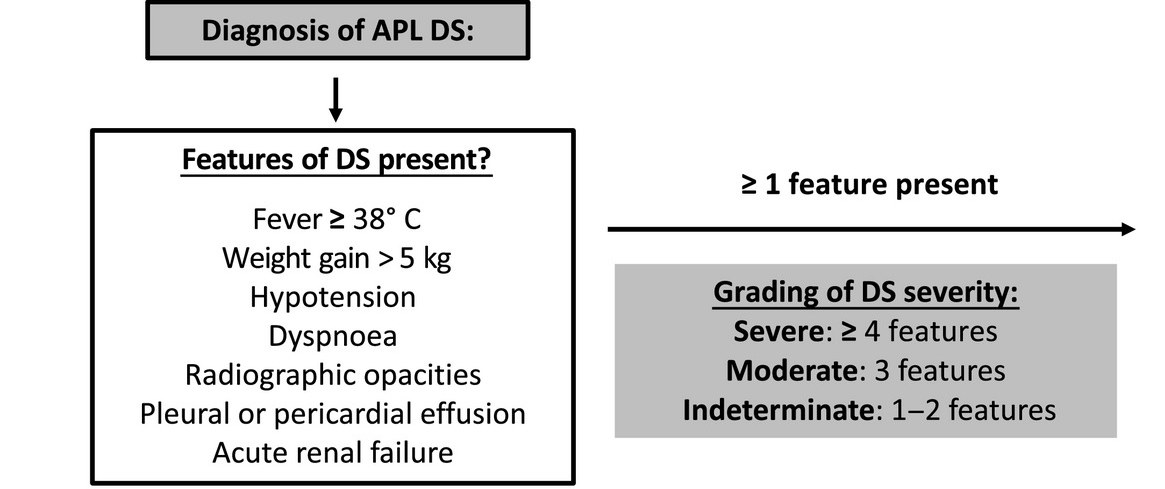
Radiographic opacities plus fluid overload — it can look exactly like ARDS.

Case 1: differentiation syndrome

Our patient got ATRA + ATO with an initial WBC of 6.4 and was started on prednisone for differentiation syndrome prophylaxis.

After 10 days, WBC rose to 35K and she developed fever + bilateral opacities + lower-extremity edema + AKI.

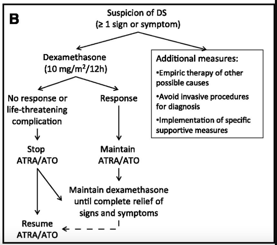
Treatment for differentiation syndrome

  • Start antibiotics and rule out other etiologies (PNA, CHF, etc.)
  • Dexamethasone 10 mg q12h
  • If no response within 24 hours, increase frequency to q6h
  • Continue high-dose dex until complete resolution of symptoms

Pearls for differentiation syndrome

Discussion

  • Do you always hold ATRA + ATO when you start dex? One, the other, or both?
  • If the patient did not develop DS but had a rising WBC — do you add hydrea to prevent it? When do you stop it?
  • When do you taper steroid prophylaxis?
Pearls for differentiation syndrome (cont.)

Discussion

Is it the same treatment for differentiation syndrome caused by IDH or menin inhibitors?

Who is Lo-Coco, and why ATRA + ATO?

What did we do prior to ATRA + ATO?

Historically, most trials came from the Italian and Spanish cooperative groups (GIMEMA and PETHEMA).

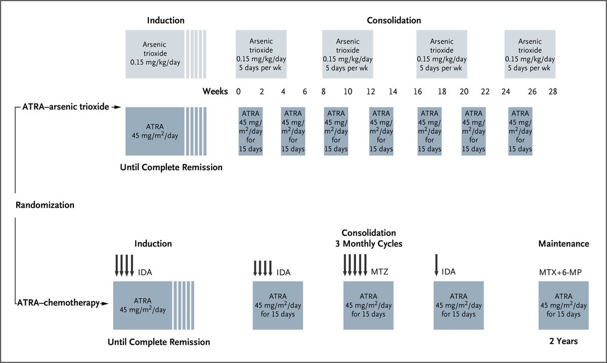
  • Induction — AIDA = ATRA + IDArubicin
    • ATRA daily; idarubicin ×4 doses every other day
  • Consolidation — ATRA + anthracycline ×3 cycles
  • Maintenance — oral 6-MP + MTX alternating with ATRA, for 2 years total

APL0406: the Lo-Coco regimen

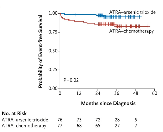
  • Noninferiority trial: low/intermediate-risk patients randomized to ATRA + ATO vs ATRA + IDA
  • ATRA + ATO arm got consolidation without maintenance
  • AIDA arm got historical induction + consolidation + maintenance
  • EFS at 2 years: 97% vs 86% (p < 0.001) — confirmed noninferiority
ATRA + ATO consolidation lasts 28 weeks

  • ATRA: 2 weeks on, 2 weeks off
  • ATO: 4 weeks on, 4 weeks off

Discussion

When do you stop ATO/ATRA for induction and discharge? What issues arise during this consolidation marathon, and how do you manage them?

APL0406: time to complete remission

They checked for morphologic CR often on trial — day 28 and then weekly.

ATRA + ATO (n=77) ATRA + chemo (n=79)
CR rate 100% 95%
Median time to CR 32 days 35 days
Range, time to CR 22–68 days 26–63 days

How often do we check marrows?

ATRA + ATO (n=77) ATRA + chemo (n=79)
CR rate 100% 95%
Median time to CR 32 days 35 days
Range, time to CR 22–68 days 26–63 days

Discussion

  • Given this data, when should we check a bone marrow biopsy? In what scenario would you do it much earlier to look for refractory disease?
  • Post-discharge, do we always keep patients on ATRA?

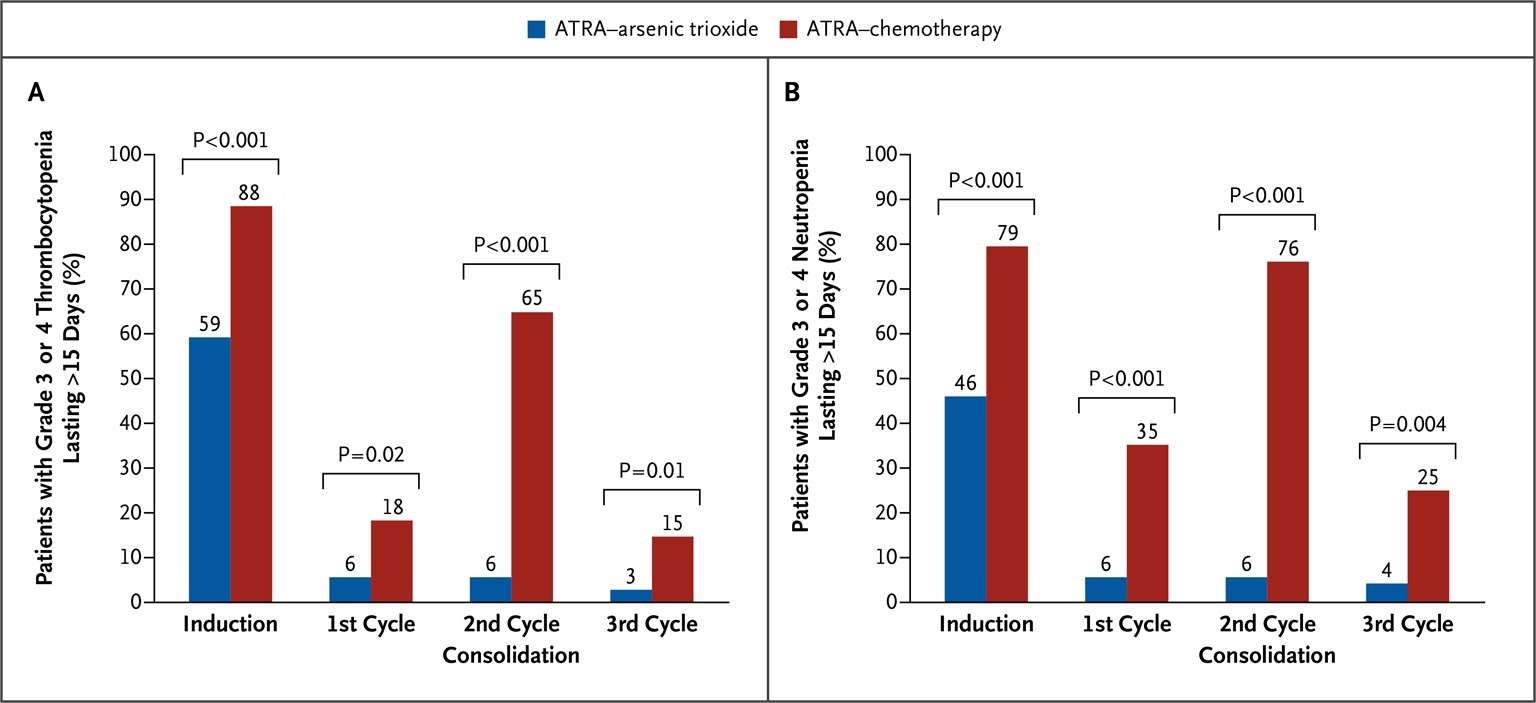
ATRA + ATO: fewer cytopenias

ATRA + ATO: the trade-off is LFTs and QTc
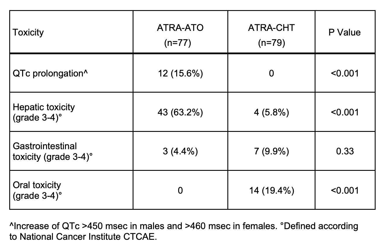
QTc prolongation 15.6% vs 0% · grade 3–4 hepatic toxicity 63.2% vs 5.8% — this is what you are monitoring for.

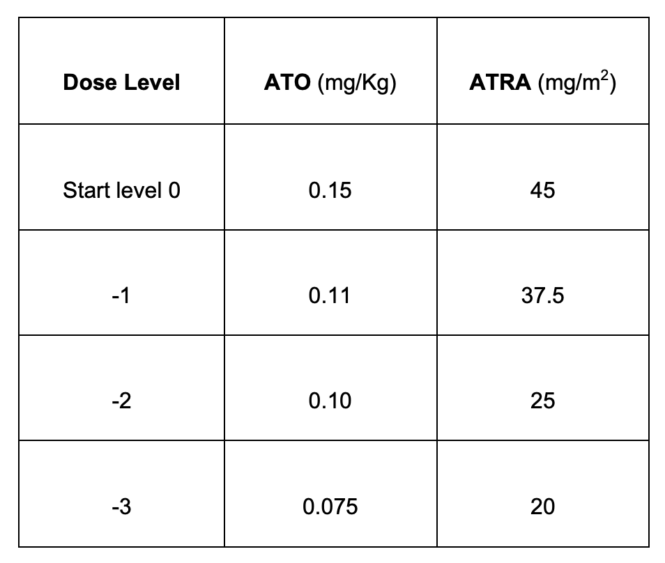
Dose reduction scheme for reference

Hepatotoxicity

  • LFTs 5× ULN (bili, ALP, or ALT)
  • Hold all therapy until <5× ULN
  • Resume at dose level −3
  • Titrate up to full dose if the trend continues down

QTc prolongation

  • >450 (men) or >460 (women)
  • Replete electrolytes and rule out other meds
  • Resume at 0.075 ×1 week → 0.11 ×1 week → full dose if no prolongation
Discussion

Hold, or dose-reduce ATO?

Case 1: putting it together

  • Step 1 — Supportive care and risk stratification: WBC < 10 → ATRA + ATO, per the Lo-Coco regimen
  • Step 2 — Watch inpatient during induction
    • Prednisone 0.5 mg/kg daily for differentiation prophylaxis
    • If WBC > 10: Lo-Coco started hydrea 500 mg daily until WBC < 10
    • Watch LFTs and QTc closely; keep Mg > 2 and K > 4
  • Step 3 — Discharge, then marrow to confirm CR (took up to 60 days on trial)

Discussion

How often do we check PML-RARA PCR during consolidation, and when should we achieve molecular CR? What about marrows?

High-risk treatment

Case 2: APL, high risk

55 y/o female presenting with significant dyspnea on 3 L O₂, with bilateral pulmonary opacities concerning for leukostasis.

Initial labs

  • WBC 80, Hgb 7.6, Plt 12
  • Differential shows 60% blasts → concerning for microgranular APL
  • INR 1.6, PTT 45, Fibrinogen 106
  • FISH positive for PML-RARA

Step 1 — Risk stratification: high risk (WBC > 10)

APL high-risk treatment

Requires induction + consolidation + 2-year maintenance.

  • Induction
    • ATRA + ATO + idarubicin, or
    • ATRA + ATO + gemtuzumab ozogamicin (CD33 antibody–drug conjugate)
  • Consolidation — ATRA + ATO; no anthracycline or GO
  • Maintenance — 2 years of 6-MP + MTX + ATRA

APML4: ATRA + ATO + IDA

  • Goal: can we add ATO to ATRA + Ida for induction and drop Ida in consolidation?
  • Phase II trial, all risk groups
  • ATRA + ATO + Ida induction
  • No anthracycline in other treatment phases
  • 2-year RFS 92% in the high-risk group

APOLLO: ATRA + ATO + IDA

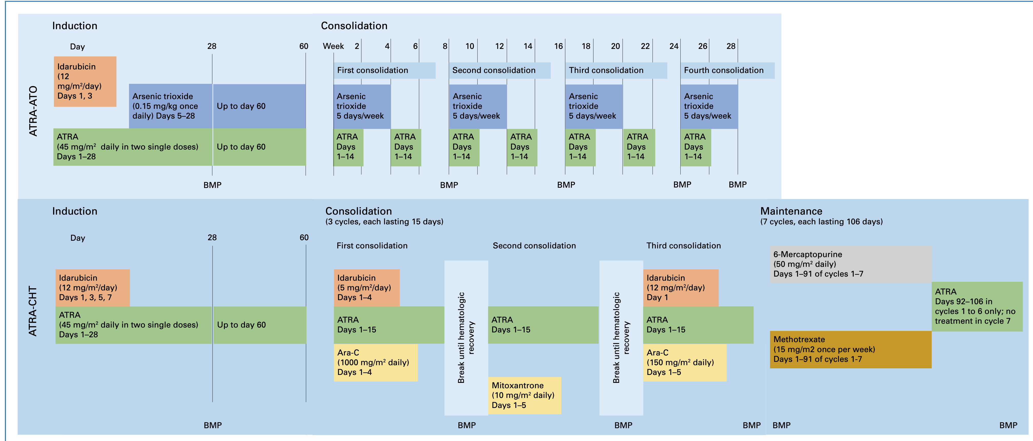
  • Open-label randomized phase III, 18–65 y/o newly diagnosed HR-APL.

  • Study was prematurely discontinued due to COVID-19

  • 131 patients were treated: 68 ATRA-ATO-AIDA and 63 ATRA-AIDA.

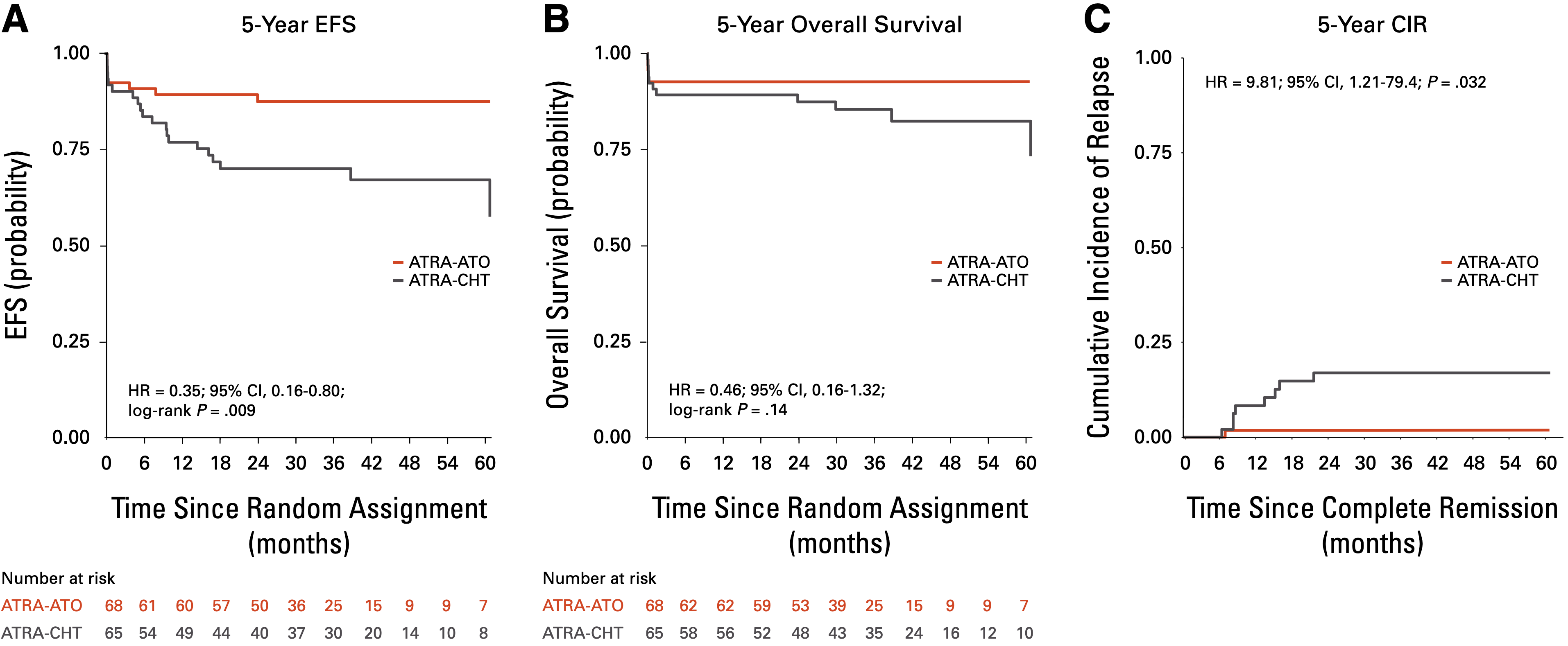
  • First-line ATRA-ATO with two initial doses of idarubicin results in superior EFS compared with conventional ATRA-CHT in high-risk APL.

  • These findings largely recapitulated the findings of APML4, despite fewer doses of IDA and the omission of a maintenance phase.
AML17: ATRA + ATO + GO

  • Phase III trial, all risk groups
  • ATRA + ATO vs ATRA + Ida
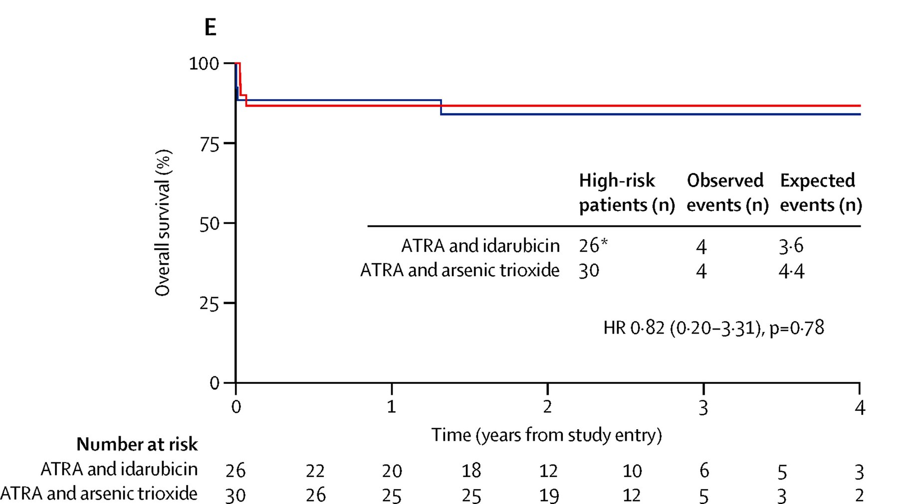
  • If high risk in the ATRA + ATO arm → add GO on day 1 only (6mg/m2)
  • No maintenance was given
  • MD Anderson phase II trials had shown efficacy of ATRA + ATO + GO
  • No difference in OS by adding GO vs idarubicin — without increased liver toxicity
ATRA-ATO-IDA vs ATRA-ATO GO

  • the Hematology Research Outcomes (HER0) Consortium
  • Multi-center Retrospective Fashion
  • 77 patients: 52 patients receiving ATRA-ATO-IDA and 25 ATRA-ATO-GO
  • Median 31 months’ follow-up, no significant differences noted in CR rates, relapse, or estimated 36 months’ survival.
  • The only significant differences were: Time to ANC and platelet recovery (shorter in ATRA-ATO/GO) and Incidence of neutropenic fever (lower in ATRA-ATO/Go)

Case 2: managing leukostasis

55 y/o female, WBC 80, 60% blasts → concerning for microgranular APL.

Discussion

How do we manage leukostasis in APL? I’ve heard that leukapheresis is contraindicated.

  • Do you give a dose or two of cytarabine?
  • Do you prefer to just start with an anthracycline-based approach?

Case 2: open questions

Discussion

  • What is your preference for management of high-risk APL?
  • What are the benefits of using GO vs idarubicin?
  • Do you keep hydrea going in this population until WBC < 10, or let the chemo do the work?
  • When do you check CNS and give prophylaxis with IT cytarabine?

Monitoring and relapse

Monitoring and relapse

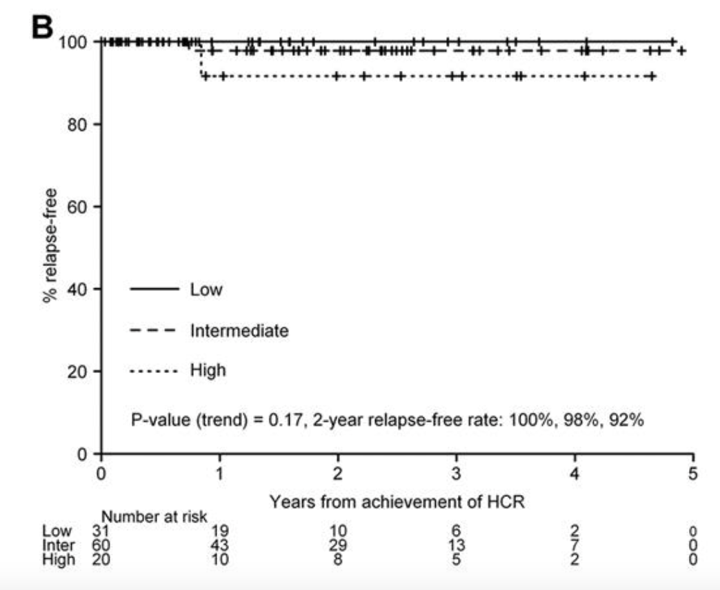
Post-Rx monitoring

  • CBC q3 months
  • PML-RARA PCR q3 months
  • Total: 2–3 years

Relapse treatment

  • ATRA + chemo, or ATRA + ATO + GO
  • Consolidate with autologous SCT

The future: oral arsenic

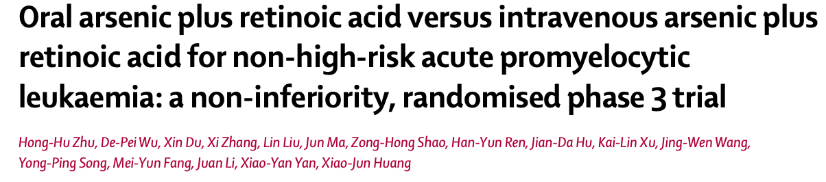
Oral arsenic (RIF)

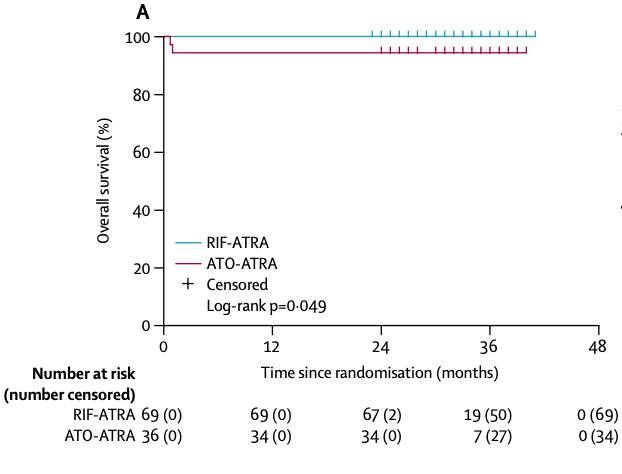
  • Oral formulation of arsenic called “RIF” — approved in Japan, not in the US
  • Phase III randomized study showed noninferiority
  • PK equivalence: Wei et al., Blood Adv 2025;9(6):1476–1484
Final thoughts

Take-home points

  • APL is a bleeding emergency — start ATRA on suspicion, before confirmation, and support aggressively
  • Diagnosis: morphology + DIC raises it, PML-RARA (FISH/PCR/karyotype) confirms it
  • Risk stratification is by presenting WBC: >10 = high risk
  • Low risk → ATRA + ATO, chemo-free, no maintenance (APL0406, Lo-Coco)
  • High risk → add idarubicin or GO, plus consolidation and no maintenance (APPOLLO)
  • Watch for differentiation syndrome, QTc prolongation, and hepatotoxicity
  • Monitor with PML-RARA PCR q3 months for 2–3 years; relapse is salvageable with autologous SCT